
Fig 3. Effect of the two catK inhibitors on AR-S staining, induced by Saos-2 cells and osteoblasts from calvaria.
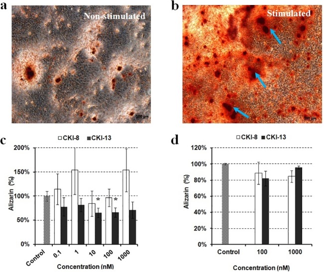
Typical example of AR-S staining on Saos-2 cells incubated during eight days a) without or b) with the addition of 50 μg mL-1 AA and 7.5 mM β-GP. The effects of control (without inhibitor in grey rectangles), CKI-8 (unfilled rectangles) and CKI-13 (black rectangles) on mineralization as indicated by their concentrations were monitored by AR-S staining on c) Saos-2 cells and d) primary osteoblasts. A t test was performed to evaluate significant differences. * p value <0.05 was regarded as statistically significant (comparison with the control).