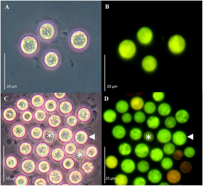
Fig 4. GFP-fusion protein fluorescence from Symbiodinium spp. cells co-incubated with Agrobacterium tumefaciens harboring a gfp-MBD fusion.
Cell cultures of S. Mf11 (A, B), and S. kawagutii (C, D) after 20 d of selection with Basta. Cells were observed under phase contrast (A and C), and under epifluorescence microscopy (B and D). Bars equal 20 μm for A and B, and 25μm for C and D.

