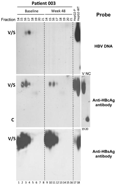
Fig. 2.
Analysis of HBV virions in human sera by CsCl density gradient fractionation. Two serum samples, at baseline (lanes 1–8) and week 48 (lanes 9–16) post-treatment, from patient 003 were fractionated by CsCl gradient centrifugation. The indicated fractions were resolved on a native agarose gel. HBV DNA, HBcAg and HBsAg were detected as described in Fig. 1. HBV virions purified from culture supernatant of HepG2 cells transfected with wild-type (WT; lane 18) HBV DNA or a mutant defective in polymerase expression (P−; lane 17) were included as controls. A virion (V) and naked capsid (NC) fraction harvested from the wild-type HBV-transfected HepG2 cell supernatant by CsCl gradient fractionation were loaded respectively in lanes 19 and 20 to show the migration of naked capsids relative to virions. Other labels are the same as in Fig. 1.

