
Figure 2. Pacing induced Ca2+ alternans.
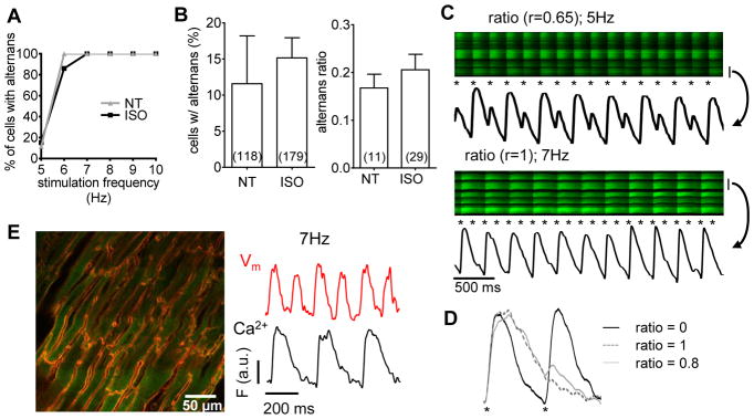
Langendorff-perfused mouse hearts under control conditions (NT) or under β-AR stimulation (ISO) were electrically stimulated at increasing frequencies and percentage of cells exhibiting alternans was assessed (A). At 5 Hz the percentage of cells with alternans and alternans ratio were unaffected by β-AR stimulation (B). Two examples of high alternans ratio are displayed. The bar at right of image indicates the cell from which signals were taken and the asterisks indicate stimuli (C). Representative transients from tissue stimulated at 5 Hz are displayed for no alternans (ratio = 0), maximal alternans (ratio = 1) and substantial alternans (r= 0.8; D). 2D image of a heart loaded with both with Ca2+ and voltage sensor (E, left). Correspondence of voltage (Vm) with Ca2+ from the same cell during 6 stimulated beats confirms action potential occurrence at missing Ca2+ transients (for r=1; E).