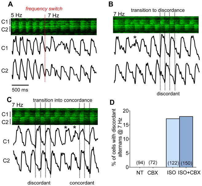
Figure 6. Transition from spatially discordant to concordant behavior.
Exemplar recording in ISO with a frequency increase from 5 to 7 Hz, showing 3 phases (A–C) with intensity profiles of two neighboring cells. The first beat at shorter BCL is reduced and quickly transitions to concordant alternans (A), which devolves into discordant alternans (B) before reverting (*) to stable concordant alternans (C). Spatially discordance was only observed in a subset of β-AR stimulated hearts and was independent of partial GJ uncoupling (D).

