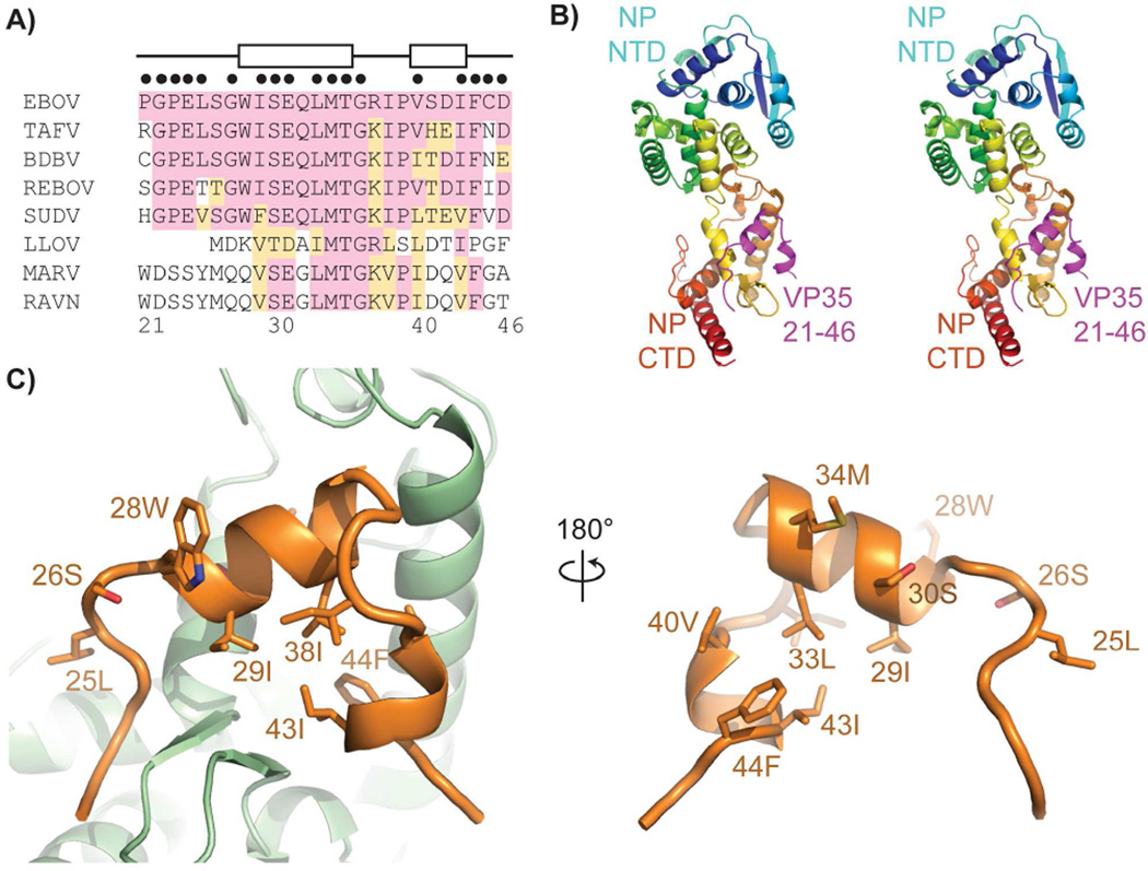
Figure 2. Structure of the NP°-VP35 complex.
A) Sequence alignment of the conserved VP35 N-terminal peptide. Amino acids sharing identity with EBOV are shaded pink, similar amino acids are shaded orange. Amino acids participating in helices as observed in the crystal structure are indicated at top by boxes. Amino acid positions within 4 Å of NP are indicated with black dots above the alignment. B) Stereoview of the NP°-VP35 crystal structure. NP is colored from the N terminus (blue) to the C terminus (red) with the VP35 peptide in magenta. C) The VP35 peptide (orange) uses a hydrophobic face and conserved amino acids to contact the NP (green). See also Figure S2 and S3 and Table S1.

