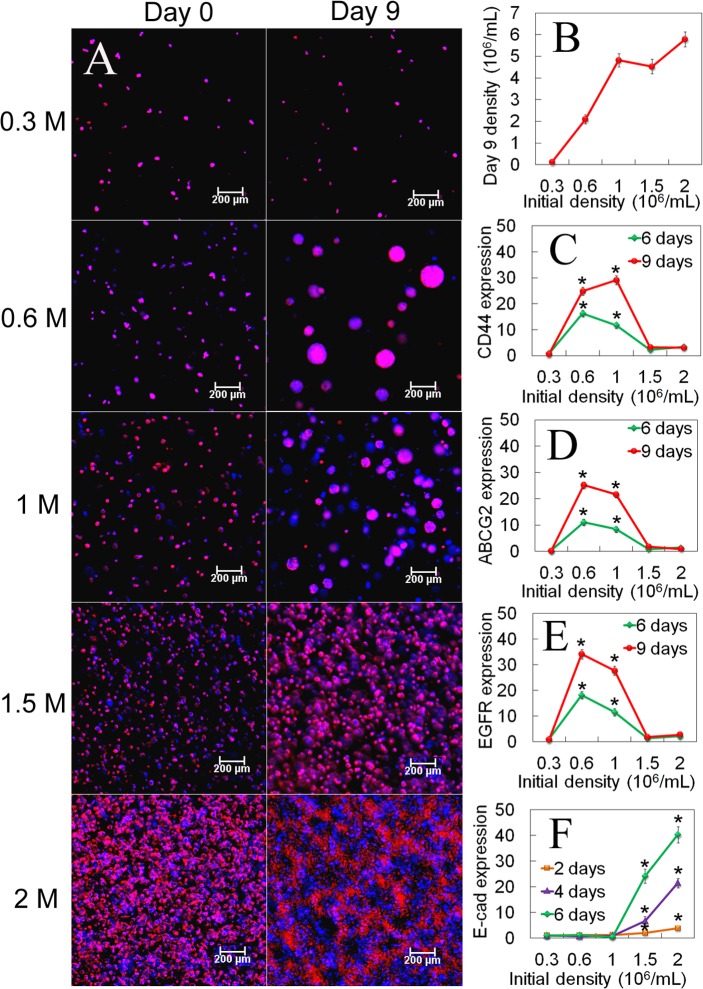
Fig 1. Dependence of tumorsphere growth on initial seeding density of MDA231 cells.
(A) DAPI (blue) and phalloidin (red) stained images of the encapsulated MDA231 cells (5 kPa gel) on day zero (left column) and after 9 days of incubation (right column) versus initial number of cells; (B) Number of encapsulated cells after 9 days of incubation as a function of initial cell number; mRNA expression of CD44 (C), ABCG2 (D), and EGFR (E) markers of the encapsulated cells after 6 and 9 days of incubation as a function of initial cell number; (F) E-cadherin mRNA expression of the encapsulated cells after 2, 4 and 6 days of incubation as a function of the initial cell number. The scale bars in (A) are 200 μm. An asterisk in (C-E) indicates a statistically higher (p<0.05) mRNA expression in the test group compared to those groups with initial cell density of 0.3x10⁶, 1.5x10⁶, and 2.0x10⁶ cells/mL at the same time point. An asterisk in (F) indicates a statistically higher E-cad expression in the test group compared to those groups with initial cell density of 0.3x10⁶, 0.6x10⁶, and 1.0x10⁶ cells/mL at the same time point. The p-values for the asterisks in (C-F) are listed in Tables A-D in S1 File. Error bars correspond to means±1 SD for n = 3.

