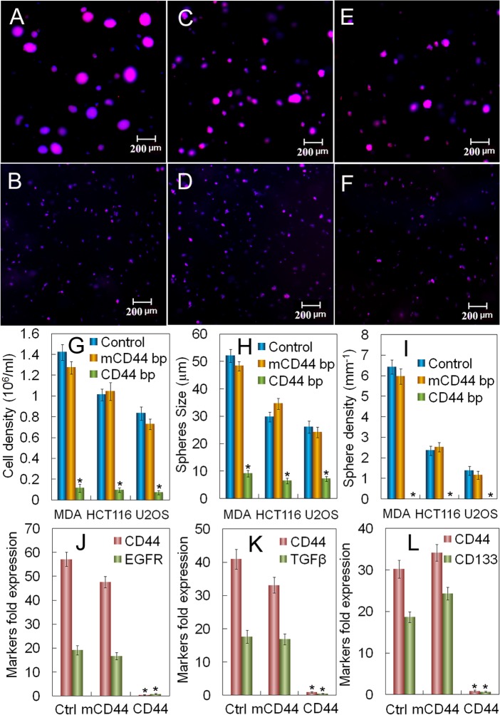
Fig 6. Dependence of tumorsphere growth on conjugation of CD44 binding peptide to the gel.
(A) DAPI (blue) and phalloidin (red) stained images of MDA231 (A+B), HCT116 (C+D), and U2OS (E+F) cells encapsulated in the un-patterned gel with optimum modulus (5 kPa for MDA231, 25 kPa for HCT116, and 50 kPa for U2OS) without CD44BP (A+C+E) and with CD44BP conjugation (B+D+F) after 9 days incubation (scale bar in A-F is 200 μm). The initial seeding density of all cell types in the gel was 0.6x10⁶ cells/mL. Cell number (G), tumorsphere size (H), and tumorsphere number (I) for MDA231, HCT116, and U2OS cells encapsulated in the gel without (blue) and with (green) conjugated CD44BP and with conjuagted mutant-CD44BP (mCD44BP, orange) after 9 days incubation. mRNA expression of CSC markers for MDA231 (J, CD44 and EGFR), HCT116 (K, CD44 and TGF-β), and U2OS (L, CD44 and CD133) encapsulated in the gel without conjugation, with mCD44BP, and with CD44BP conjugation. An Asterisk in (G-L) indicates a statistically lower cell number, sphere number and size, and marker expression for the test group compared to those groups without CD44BP conjugation and with mutant CD44BP conjugation of the gel for a given cell type. The p-values for the asterisks in (G-L) are listed in Tables A-F in S2 File. Error bars correspond to mean±1 SD for n = 3.

