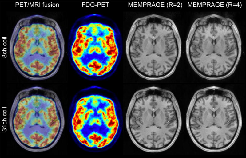
Figure 10.

Simultaneously acquired PET ([18F]FDG) and MRI in a human subject shown as a fused image (left). Artifact-free PET images (left center) demonstrate an accurate implementation of the 31-channel coil attenuation correction. MR images obtained with the 8-channel and 31-channel coil show T1 anatomy (MEMPRAGE at acceleration R=2 (center right) and R=4 (right)) and the superior g-factor and SNR of the 31-channel coil.
