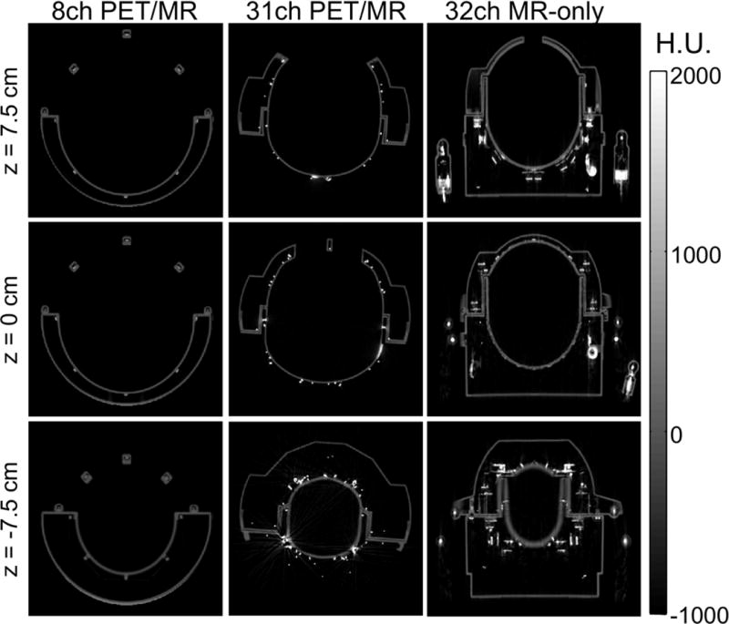
Figure 7.

Axial CT scans for z-positions −7.5 cm, 0 cm and 7.5 cm of the 8-channel PET/MR, the 31-channel PET/MR and a standard MR-only 32-channel coil. While the 8-channel coil shows the least material in the PET FOV, the 32-channel MR-only coil shows attenuating coil components, such as the plugs, preamplifiers and cable traps. Compared to these two coils, the 31-channel PET/MR coil shows more metal due to the higher number of coils but does not have large attenuating coil components in the PET FOV.
