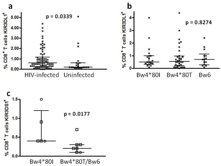
Figure 1.

Distributions of percentage CD8+ T cells expressing KIR3DL1 or KIR3DS1 in different HIV-infected and uninfected subgroups. The percentage of CD8+ T cells expressing KIR3DL1 was compared between HIV-infected and uninfected KIR3DL1 homozygous subjects (a) and between subgroups of the HIV-infected individuals distinguished by HLA-Bw4*80I, Bw4*80T or Bw6 status (b). The percentage of CD8+ T cells expressing KIR3DS1 was compared between KIR3DS1 homozygous subjects expressing Bw4*80I or not with different symbols used to distinguish HIV-infected (□) from uninfected (○) individuals (c).
