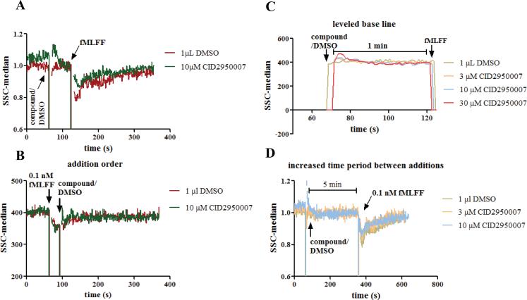
Figure 2.
Right angle side scatter assay development. (A) Compound or DMSO was added to PMN cells. When the right angle side scatter baseline was stabilized one minute later, 0.1 nM fMLFF was added. The compound showed different effects from DMSO. (B) fMLFF was added before compound or DMSO. No apparent difference was observed between compound and DMSO. (C) Expanded view of the right angle side scatter reading between compound/DMSO and fMLFF additions. The baseline reading had stabilized before fMLFF addition. (D) fMLFF was added 5 min after the compound or DMSO. The dose-dependent effects of CID2950007 are comparable to that when fMLFF was added 1 min later. Experiments were conducted using PMNs from two healthy donors and repetition numbers are 2 and 3, respectively. Representative curves are shown. For (A) and (D), the right angle side scatter reading was normalized by being divided with an average of the readings before the fMLFF addition. Ten time points were averaged.

