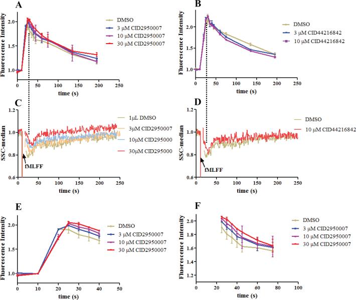
Figure 3.
Fluorescent phalloidin staining assay for comparison. (A) Fluorescence readings of PMNs treated with CID2950007 or DMSO followed by fMLFF. (B) As in (A) except the compound is CID44216842 which facilitated depolymerization. (C) Right angle side scatter readings of PMNs as treated in (A). The changes of readings were coincident with that in (A) without distinct resolution. (D) Right angle side scatter readings of PMNs as treated in (B). The changes of readings were coincident with that in (B) without distinct resolution. (E) Expanded view of (A) for the first 40 s. CID2950007 dose dependently delayed the polymerization process. The slope of the red line was smaller than that of the purple, blue and grey lines in order. (F) Expanded view of (A) after the first 25 s. The depolymerization process was also dose dependently delayed. Both the fluorescence and the side scatter readings were normalized by being divided with an average of the readings before the fMLFF addition. For experiments shown in Figure 3B and D, PMNs from two healthy donors were used and repetition numbers are 2 and 3, respectively, while for other experiments, PMNs from four healthy donors were used and repetition numbers are 2, 3, 3 and 3, respectively.

