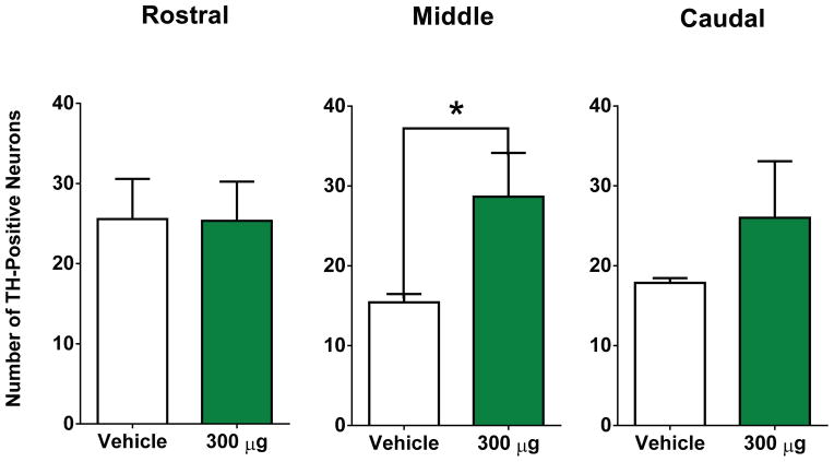
Figure 7. The effects of repeated intranasal administration of DNSP-11 on TH+ dopaminergic neurons in the SNpc and fiber area of the SNpr in 6-OHDA unilaterally lesioned rats.
(A–C) The total number of TH+ neurons were counted from three sections of the lesioned SNpc after repeated intranasal administration of either vehicle or DNSP-11: Rostral (t(11) = 0.2714, p = 0.7911), (A–D) Middle (t(11) = 2.703, p = 0.0205), and Caudal (t(11) = 1.223, p = 0.2468). There was no significant difference found between DNSP-11 treated rats or vehicle in the rostral or caudal regions of the SNpc (mean ± SEM, vehicle: 23 ± 6; DNSP-11: 25 ± 5) and (mean ± SEM, vehicle: 18 ± 1; DNSP-11: 26 ± 7). (B) There was a significant increase observed in the average number of TH+ positive neurons in DNSP-11 treated rats compared to vehicle (*p < 0.05) in the middle region of the lesioned SNpc (mean ± SEM, vehicle: 13 ± 2; DNSP-11: 29 ± 5). The data were analyzed by a two-tailed unpaired t test. Data are shown as mean ± SEM (n = 7 vehicle, n = 6 DNSP-11).

