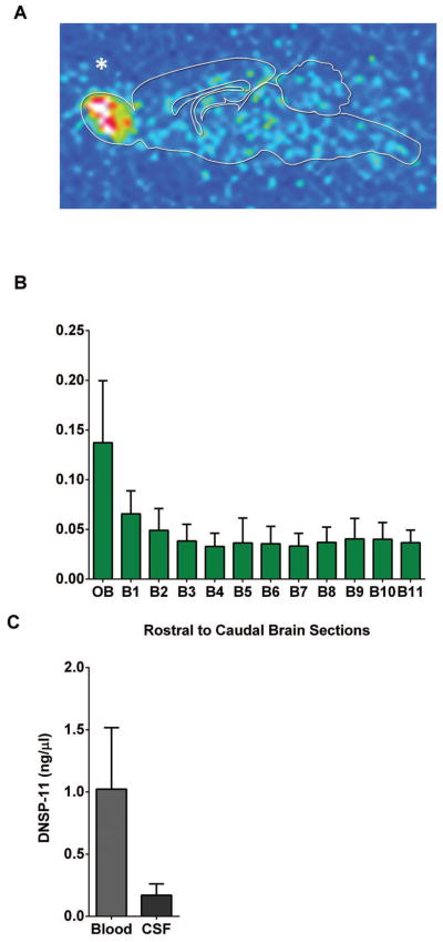
Figure 8. Tracking of a one-time 125I-labeled DNSP-11 dose 60 minutes after intranasal administration.
Normal F344 rats were given a one-time intranasal dose of 125I-labeled DNSP-11, at 60 minutes blood (500 μl) and cerebrospinal fluid (100–120 μl) were collected from each rat and processed by gamma counting (n=3) and autoradiography (n=1). (A) Representative sagittal brain section (0.5 mm thick), exposed for 21 days on a GE phosphor screen. A qualitative increase in radioactive signal was found in the olfactory bulbs 60 minutes post intranasal administration and diffusely throughout the brain. (B) Data are shown as the normalized DNSP-11 concentrations (ng or μl/mg of wet sample weight) as analyzed by gamma counting at 60 minutes. (C) Blood and CSF; the visible increase in radioactive signal found in the olfactory bulbs at 60 minutes is consistent with autoradiography. Data are presented as the normalized DNSP-11 concentrations (ng or μl/mg). Rats treated with vehicle only were found to have CPM lower then background levels < 50 (data not pictured). * Denotes the olfactory bulb of a representative sagittal section of the midbrain after autoradiography analysis.

