Figure 3.
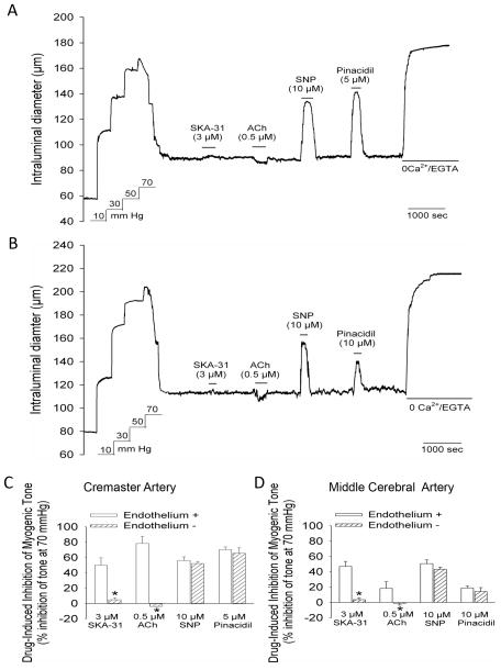
Endothelium denudation prevents SKA-31 and ACh-evoked vasodilation in cremaster and middle cerebral arteries. Panel A shows a representative tracing depicting the vasodilatory responses to 3 μM SKA-31, 0.5 μM ACh, 10 μM SNP and 5 μM pinacidil in a cremaster artery in which an air bubble had been passed through the vessel lumen prior to the development of myogenic tone. The representative tracing in panel B shows vasodilatory responses to the same vasodilators in a middle cerebral artery in which the endothelium had been disrupted by the introduction of an air bubble. The histograms in panels C and D depict the magnitude of dilator-evoked inhibition of myogenic tone in cremaster (panel C) and middle cerebral arteries (panel D) with intact endothelium (+) or physically disrupted endothelium (-). Vasodilatory responses to SKA-31, ACh, SNP and pinacidil in endothelium-intact vessels were derived from other experiments reported in this study; for intact cremaster arteries, n = 6–14, for intact middle cerebral arteries, n = 4–7. For both endothelium-denuded cremaster and middle cerebral arteries, average values were calculated from 4 vessels of each type. Data are expressed as means ± SEM. The asterisk (*) indicates a statistically significant difference in the magnitude of inhibition compared with the intact endothelium condition, as determined by an unpaired Student’s t-test (P < 0.05).

