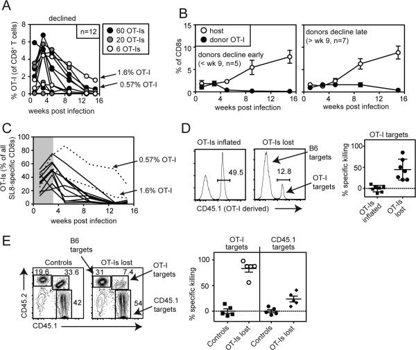
Figure 7.
Rejection of transferred OT-Is allows host-derived SIINFEKL-specific T cells to inflate. A) 60, 20 or 6 DP OT-I T cells were transferred into B6 (CD45.2+) recipients before infection with MCMV-GFP-SL8. Shown is the frequency of DP OT-Is in the peripheral blood in mice in which OT-Is were lost over time. Arrows indicate two mice in which OT-Is declined but still comprised 1.6% and 0.57% of all CD8+ T cells in the peripheral blood at the final time point. B) Inflation of donor OT-Is and host-derived SIINFEKL-specific T cells in the peripheral blood is shown for the mice shown in “A”. Animals were grouped into those in which OT-Is were lost by week 9 (left graph) or declined but comprised more than 0.3% of all CD8 T cells at least 9 weeks post infection (right graph). C) The total SIINFEKL-specific response was measured by IFN-γ production from peripheral blood T cells and the percentage of all responding cells that were donor-derived OT-Is was plotted over time. The shaded area visually denotes the early selection of OT-Is relative to host-derived SIINFEKL-specific T cells. Data for A-C are combined from the three independent experiments. D) In vivo killing assays were conducted as in Figure 6 except that mice received a mixture of CFSE-labeled B6 splenocytes (CD45.2+ controls) and DP OT-I splenocytes. Specific killing was calculated for each animal (n=7). Data are combined from two independent experiments. E) As in “D” except that mice received a CFSE-labeled mixture of B6 splenocytes (CD45.2+ controls), B6.SJL splenocytes (CD45.1+ nontransgenic targets), or DP OT-I splenocytes. Shown are representative FACS plots and specific killing of target populations from one experiment (n=5).

