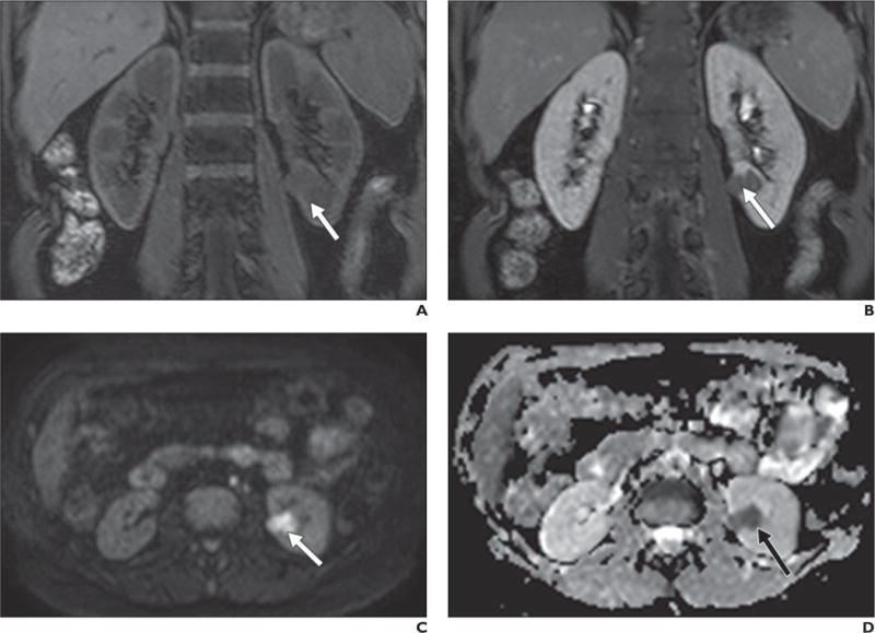
Fig. 10.

50-year-old woman with renal cell carcinoma (RCC).
A–D, Coronal T1-weighted 3D spoiled gradient-echo images obtained before (A) and after (B) gadolinium administration show left renal mass (arrow) with subtle enhancement. Lesion was iso- to slightly hypointense to kidney on low-b-value images (not shown) but is hyperintense compared with renal parenchyma on high-b-value diffusion-weighted image (C) and hypointense on apparent diffusion coefficient map (D). Findings are consistent with restricted diffusion, confirming solid neoplasm in this context and papillary RCC was confirmed at histopathology after partial nephrectomy.
