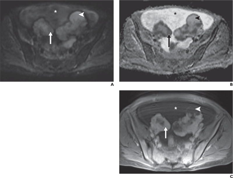
Fig. 12.

49-year-old woman with bilateral ovarian metastases from gastric adenocarcinoma (Krukenberg tumors).
A–C, Diffusion-weighted image with b800 (A), apparent diffusion coefficient (ADC) map (B), and contrast-enhanced T1-weighted spoiled gradient-echo image (C) show solid enhancing components (compared with unenhanced image, not shown) in ovarian masses (arrow), high signal intensity on b800 images, and low signal intensity on ADC map. Surrounding ascites (asterisk) has intermediate signal intensity on high-b-value image and high signal intensity on ADC map, consistent with T2 shine-through on b800 images. Cystic portions of ovarian masses (arrowheads) also show high signal intensity on high-b-value image and ADC map but are not as hyperintense as surrounding ascites on ADC map, suggesting more complex (e.g., proteinaceous or hemorrhagic) fluid.
