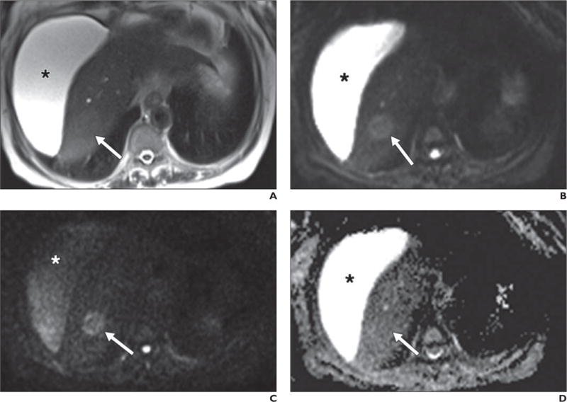
Fig. 4.

63-year-old woman with cirrhosis and new mass identified on ultrasound (not shown).
A–D, Lesion (arrow) nearly isointense to background liver on axial T2-weighted single-shot fast spin-echo image (A) is well shown on low-b-value diffusion-weighted (b = 50) image (B). Retained signal on high-b-value (b800) image (C) suggests solid lesion, which as new finding in setting of cirrhosis is worrisome for hepatocellular carcinoma. Note abdominal ascites (asterisk), which decreases in signal intensity on high-b-value images, suggestive of simple fluid. This fluid was bright on ADC map (D). Dynamic imaging was not performed due to contraindication to gadolinium. This example also shows potential inconspicuity of solid lesion on ADC map (D) on background of cirrhotic liver.
