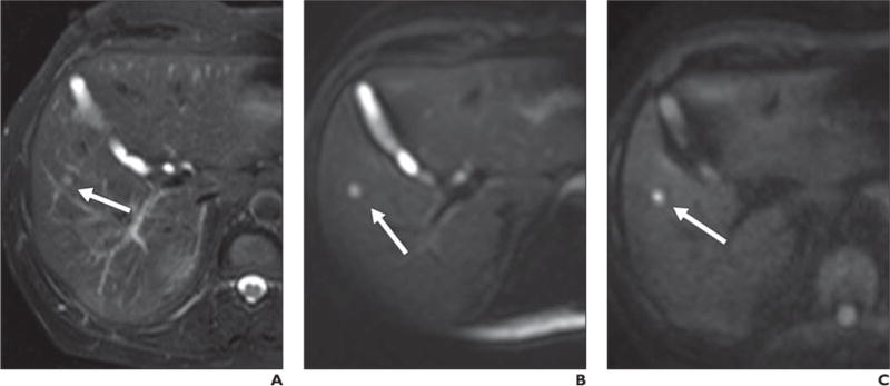
Fig. 5.

69-year-old man with lung cancer.
A, Axial T2-weighted fat-saturated single-shot fast spin-echo image shows small hyperintense lesion (arrow), which is difficult to differentiate from adjacent vessels.
B, Low-b-value (b = 50) diffusion-weighted image shows lesion (arrow) with far more conspicuity than T2-weighted sequence due to cancellation of intravascular signal.
C, Lesion (arrow) retains signal intensity on high-b-value (b800) image and is consistent with restriction in solid lesion. This patient could not receive contrast agent and diffusion-weighted imaging provided best opportunity to detect this lesion, suspicious for metastatic focus.
