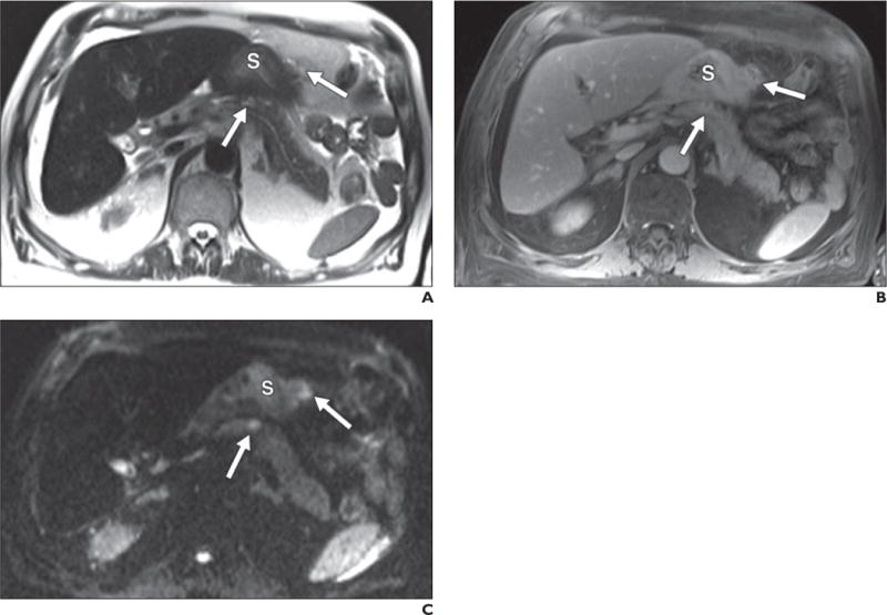
Fig. 7.

67-year-old man with peritoneal metastatic deposits (arrows) related to gastric adenocarcinoma.
A–C, Peritoneal disease is subtle on T2-weighted single-shot fast spin-echo (A) and gadolinium-enhanced T1-weighted spoiled gradient-echo (B) images but most conspicuous on diffusion-weighted b400 image (C) on which it is seen as foci of bright signal intensity. S = stomach.
