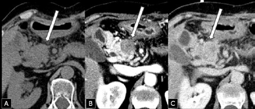
Figure 1.

Computed tomography scan after six years from primary distal pancreatectomy for ductal adenocarcinoma showing an enlargement of the pancreatic head containing a mass (arrow) measuring 28x23 mm, isodense at baseline examination (A), relatively hypodense after rapid contrast medium injection (arterial phase) (B), and isodense in the portal phase (C).
