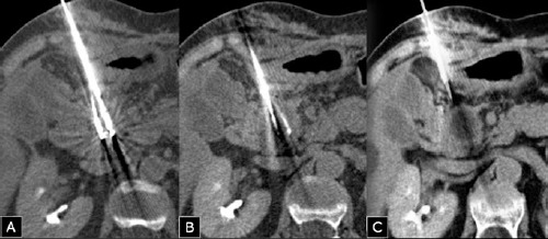
Figure 3.

Two cryoprobes (Icesed®) with active tip positioned within the tumor were inserted under computed tomography (CT)-guidance with the patient in the supine position (A, B). The CT scan performed during the freezing phase showed an hypodense area located in the site of the tumor, corresponding to the ice ball (C).
