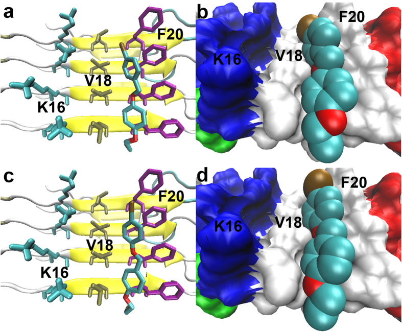
Figure 2. Lowest energy docked conformations of 7a (a,b) and 12a (c,d) packed against the hydrophobic Val18_Phe20 channel located on the flat surface of Aβ fibers (PDB ID: 2LMO).
In the right-hand panels, the molecular surface of the Aβ fiber is represented with an opaque surface and the atomic radii of ligands with solid spheres to more vividly illustrate the tight binding of the ligands to the hydrophobic groove.

