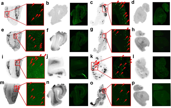
Figure 5. In vitro autoradiography of [125I]7a (a–d), [18F]7a (e–h), [125I]12a (i–l) and [18F]12a (m–p).
(a,e,i and m) AD human brain sections, female, 64 years old; (b,f,j and n) normal human brain sections, male, 74 years old; (c,g,k and o) Tg mouse brain sections, APPswe/PSEN1, 11 months old; (d,h,l and k) wild-type mouse brain sections, C57BL6, 11 months old. Fluorescence staining using ThS on the right panel of each image confirmed the location and distribution of plaques on the same sections.

