
Figure 5. Proliferation of human bone marrow-derived mesenchymal stem cells on PLGA/PEG-alginate scaffolds.
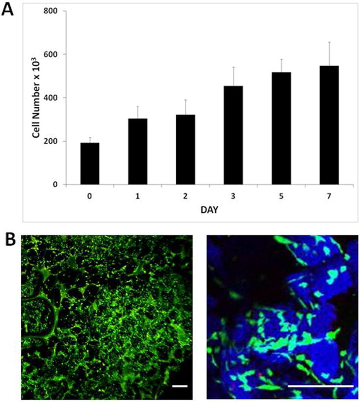
(A) Number of viable human bone marrow mesenchymal stem cells (hBM-MSCs) on 40% PLGA/PEG-60% alginate as measured by the Prestoblue metabolic activity assay over 7 days in culture. Error bars represent standard deviation. (B) Representative images of hBM-MSC-seeded 40% PLGA/PEG-60% alginate scaffold 3 days post-seeding following Live/Dead staining. PLGA/PEG particles rendered in blue using Leica LCS confocal macroscope software in order to visualise the particles (blue) and pore spaces between particles (black) Size bar = 200μm.