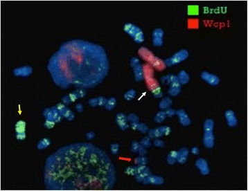
Fig. 3.

Late replication assay. Fluorescent BrdU assay, combined with WCP1 probe FISH, shows that the normal X chromosome was late replicating (yellow arrow). The late replication also interested the region Xq21-q22 translocated to der(1) (white arrow). Red arrow points to der(X)
