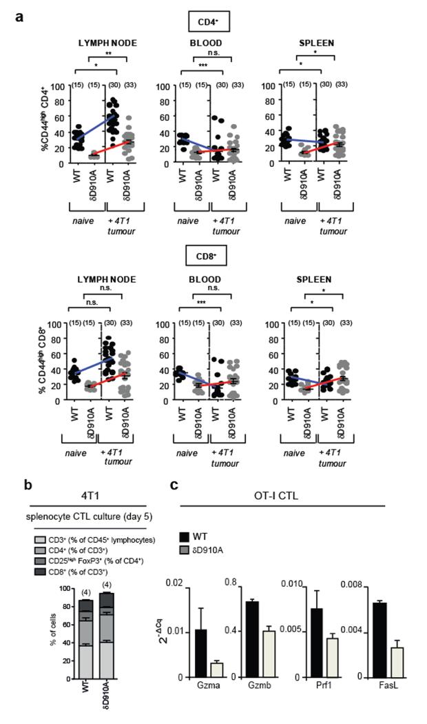
Extended Data Figure 1. Impact of p110δ inactivation on CD4 and CD8 T cells in mice with 4T1 or EL4 tumours.
a, Levels of CD44highCD4+ and CD44highCD8+ T cells in the indicated immune compartments of naive and 4T1 tumour-bearing on day 26 after inoculation in WT or δD910A mice. b, Distribution of cells on day 5 of culture of splenocytes, isolated from 4T1 tumour-bearing WT and δD910A mice 21 days after inoculation, in the presence of mitomycin-treated 4T1 cells. c, Gene expression in CTLs derived from splenocytes from WT and δD910A OT-I mice, cultured in the presence of SIINFEKL OVA peptide and IL2. GzmA, granzyme A; GzmB, granzyme B, Prf1, perforin and (FasL or CD95L) Fas ligand. Expression levels are presented relative to β2-microglobulin. a-b, Statistically significant differences are indicated by * (P < 0.05) or ** (P < 0.01), as determined by the non-parametric Mann-Whitney t test. Between brackets: number of mice used per experiment. Each dot represents an individual mouse.

