
Fig. 6.
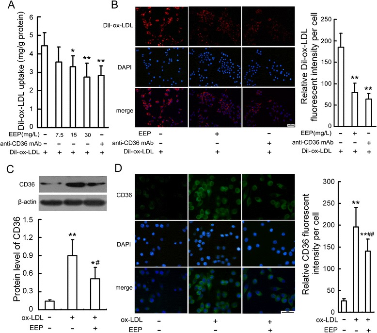
EEP attenuates ox-LDL uptake and CD36 upregulation in RAW264.7 cells. a Dil-ox-LDL fluorescence intensity in cells preincubated with EEP at the indicated concentrations or anti-CD36 mAb (2 mg/L) for 1 h and then treated with Dil-ox-LDL (50 mg/L) for 6 h. b Fluorescence microscopy showed Dil-ox-LDL uptake by RAW264.7 cells preincubated with EEP (30 mg/L) or anti-CD36 mAb (2 mg/L) for 1 h and then treated with Dil-ox-LDL (50 mg/L) for 6 h. Red staining denotes Dil-ox-LDL fluorescence and the blue staining denotes nuclei visualized by DAPI (Scale bar = 20 μm). c and d Protein expression of CD36 in cells pretreated with EEP (30 mg/L) in the presence of ox-LDL (100 mg/L) for 24 h were evaluated by Western blot and immunofluorescence assay, respectively. Representative fluorescent images are shown. Green, CD36 visualized by FITC labeling; blue, nuclei visualized by DAPI. Scale bar = 20 μm. Data are expressed as the mean ± SEM of at least three independent experiments. *P < 0.05, **P < 0.01 versus vehicle-treated control; # P < 0.05, ## P < 0.01 versus ox-LDL treatment