Figure 3.

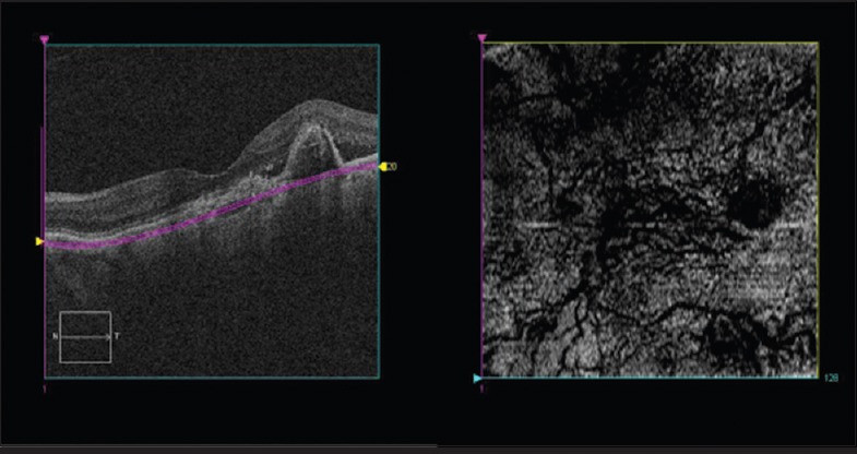
En-face image (right panel) of an eye with polypoidal choroidal vasculopathy showing abnormal vascular network just below the level of retinal pigment epithelium as shown on cross-sectional scan (left panel)

En-face image (right panel) of an eye with polypoidal choroidal vasculopathy showing abnormal vascular network just below the level of retinal pigment epithelium as shown on cross-sectional scan (left panel)