
Fig. 1.
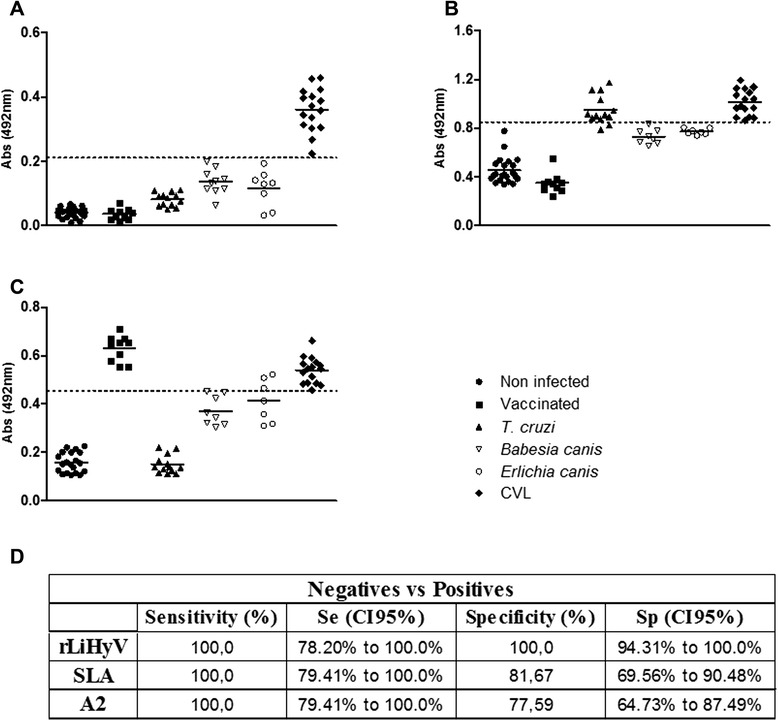
Diagnostic performance of LiHyV protein. ELISA assays were performed using sera samples of dogs with visceral leishmaniasis (n = 16), non-infected dogs living in an endemic area of leishmaniasis (n = 20), Leish-Tec®-vaccinated animals (n = 10) and animals experimentally infected by Trypanosoma cruzi (n = 13), Ehrlichia canis (n = 7) or Babesia canis (n = 7). The rLiHyV (a), SLA L. infantum (b), and rA2 (c) were used as antigens. The optical density (O.D.) values of each individual serum are shown. The cut-off values (dotted line) for negative and positive sample discrimination were calculated using the mean ± three times the standard deviation of all negative samples. The sensitivity and specificity values of the antigens were calculated and are also showed (d)