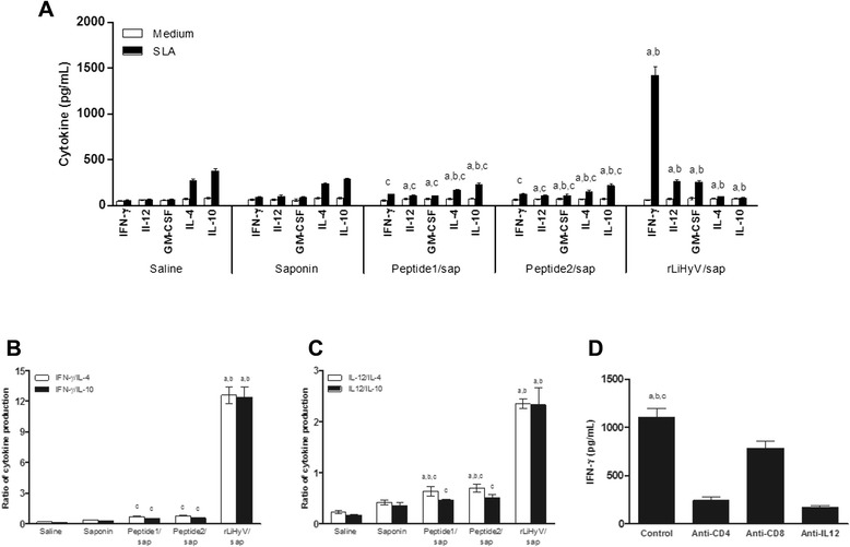
Fig. 5.

Analysis of cellular response and involvement of CD4+ and CD8+ T cells in the IFN-γ production after L. infantum infection. Spleen cells of mice were obtained 10 weeks after challenge, and were in vitro stimulated with SLA (25 μg per mL), for 48 h at 37 °C in 5 % CO2. IFN-γ, IL-12, GM-CSF, IL-4 and IL-10 levels were measured by a capture ELISA (a). The ratios between IFN-γ/IL-10 and IFN-γ/IL-4 levels (b), as well as between IL-12/IL-10 and IL-12/IL-4 levels (c) were calculated and also shown. Statistically significant differences in relation to the saline, saponin or rLiHyV/saponin groups were observed (a, b and c letters, respectively; P < 0.05). The involvement of CD4+ and CD8+ T cells in the IFN-γ production in the rLiHyV/saponin group was evaluated (d). Statistically significant differences in relation to the use of anti-IL-12, anti-CD4 or anti-CD8 monoclonal antibodies were observed (a, b and c letters, respectively; P < 0.05). Bars represent the mean ± standard deviation of the groups
