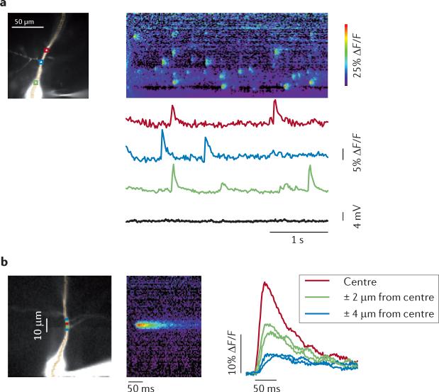
Figure 4. Spontaneous Ca2+ release events occur in localized regions of the dendrites of hippocampal pyramidal neurons.
a | The left image shows a CA1 pyramidal neuron in an acute rat hippocampal slice filled with 100 μM Oregon Green BAPTA1. Three regions of interest (ROIs) are marked with coloured boxes. The string of pixels along the dendrite indicates the location of the line scan image to the right. In normal artificial cerebral spinal fluid, spontaneous increases in intracellular calcium concentration ([Ca2+]i) were detected asynchronously at the three locations. There was no corresponding change in membrane potential. The pseudocolour line scan image (right panel) shows the increases in [Ca2+]i at all locations along the dendrite. b | The left image shows averaged records of event signals from nearby locations on a pyramidal cell dendrite. The data were recorded at 500 Hz, and spontaneous event signals from 11 events at the same location were aligned at the starting time of the rising phase of the fluorescence transient. The pseudocolour image shows the line scan of the averaged signal. The traces show the signal at the centre and neighbouring (± 2 μm and ± 4 μm from centre) locations. The data are consistent with Ca2+ release at the centre and Ca2+ diffusion to nearby locations. ΔF/F, relative change in fluorescence. Figure is adapted, with permission, from REF. 104. © (2009) Society for Neuroscience.

