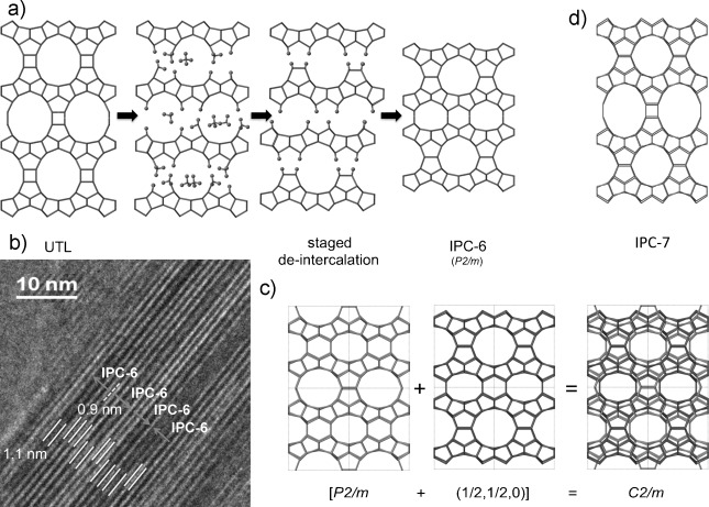
Figure 4.

Staged structure of IPC-6. a) Formation of IPC-6 based on a staged de-intercalation mechanism. b) Representative TEM image showing the two different lattice fringe spacings (1.1 nm and 0.9 nm) and how two different settings of the IPC-6 unit cell can be used to describe the overall structure of the particles. c) Two orientations of IPC-6 give an average structure that has the same symmetry as the PXRD pattern. d) Structure of IPC-7.
