. 2015 Jun 25;4:e06821. doi: 10.7554/eLife.06821

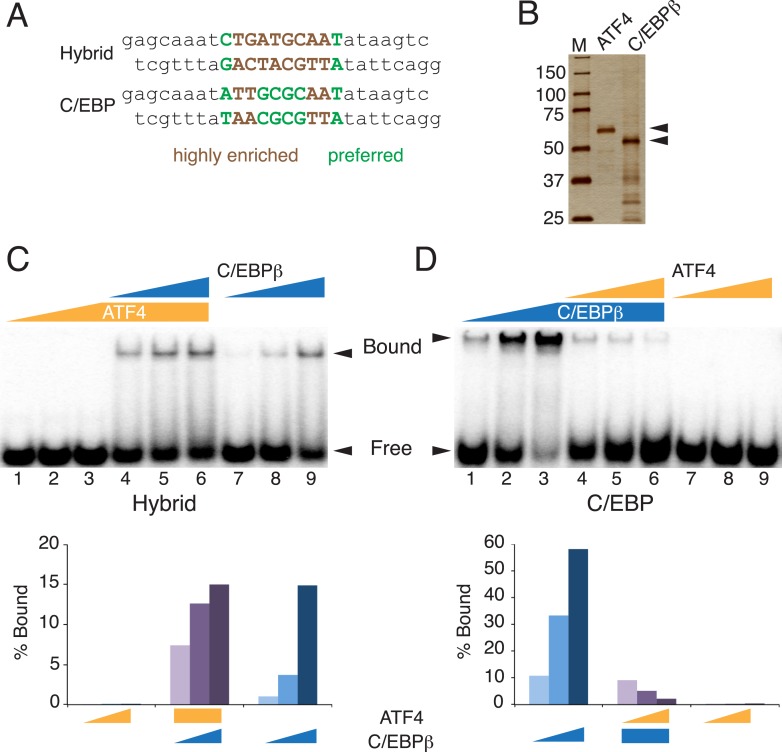
Figure 5. Distinct DNA-binding activities for ATF4, C/EBPβ and the C/EBPβ-ATF4 heterodimer.

(A) EMSA probe design for the hybrid and C/EBP sequences. Motifs are capitalized. (B) Silver-stained SDS-PAGE of recombinant his-tagged ATF4 and C/EBPβ. Arrowheads indicate the expected MWs for the tagged constructs. (C and D) EMSA titration of ATF4 (10, 30 and 90 μM) and C/EBPβ (100, 300 and 900 nM) with 0.25 nM of either the hybrid (C) or C/EBP (D) radiolabeled probe. Bar graphs show phosphoimager-based quantification of bound complexes relative to free probe.

DOI: http://dx.doi.org/10.7554/eLife.06821.011

Figure 5.

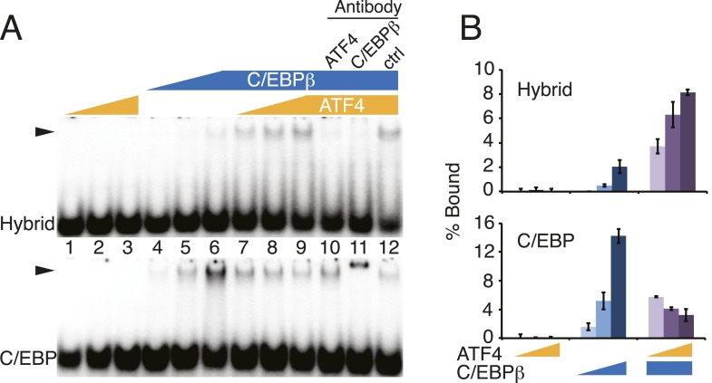
Figure 5—figure supplement 1. DNA-binding activities for ATF4, C/EBPβ and the C/EBPβ-ATF4 heterodimer.

Figure 5—figure supplement 1.

(A) EMSA titration of ATF4 (2, 6 and 18 μM) and C/EBPβ (50, 150 and 450 nM) with 0.25 nM of either the hybrid (upper) or C/EBP (lower) radiolabeled probe. Antibody controls show a supershift for the C/EBPβ homodimer, however are inhibitory for DNA binding by the C/EBPβ-ATF4 heterodimer. Antibodies were added to recombinant proteins prior to the addition of probe. Arrowheads indicate gel–shift complexes. (B) Bar graphs quantifying probe binding from two independent EMSA experiments; error bars depict the data range.