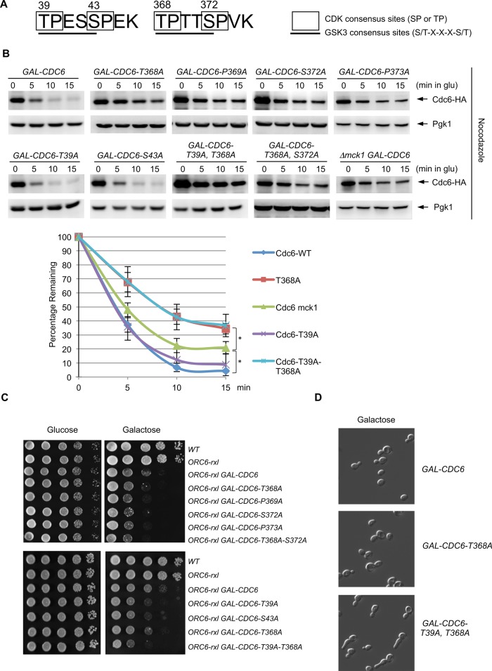
FIGURE 1:
Analysis of GSK-3 consensus sites in Cdc6. (A) Cdc6 contains two GSK-3 consensus sites, which overlap with two CDK consensus sites. (B) GAL-CDC6-HA strains with various mutations (T368A, P369A, S372A, P373A, T39A, S43A, T39A-T368A, and T368A-S372A) were expressed with galactose-containing medium for 2 h and then blocked with nocodazole for 2 h. Cdc6 expression was then suppressed by adding glucose. Protein extracts were collected every 5 min and subjected to Western blot analysis to observe Cdc6-HA. Pgk1 was used as a loading control. GAL-CDC6-HA in mck1-deletion cells was examined using the same method. Western blotting images for WT, Δmck1, CDC6-T39A, CDC6-T368A, and CDC6-T39A,T368A were quantified. Percentage of Cdc6 protein remaining relative to time zero is shown. Results are the average of three independent experiment, and error bars indicate SD. *p < 0.05. (C) Strains with the indicated genotypes were serially diluted 10-fold, plated on yeast extract/peptone/dextrose or yeast extract/peptone/galactose plates, and incubated at 30°C for 2 d. (D) GAL-CDC6, GAL-CDC6-T368A, or GAL-CDC6-T39A-T368A was grown in raffinose-containing medium first. Cdc6p was expressed with galactose for 3 h.

