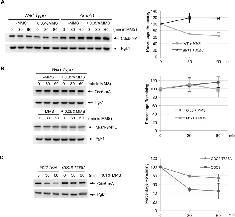
FIGURE 4:
DNA damage triggers Cdc6 degradation in Mck1-dependent manner. (A) CDC6-prA or Δmck1 CDC6-prA cells were incubated in yeast extract/peptone/dextrose (YEPD) medium to log phase, and then MMS was added (0.05% final). Protein extracts were made at 0-, 30-, or 60-min incubation and subjected to Western blotting to visualize endogenous Cdc6-prA. Pgk1 was used as a loading control. The same experiment was repeated three times to quantify Cdc6 protein levels. Average percentage of Cdc6p remaining is shown. Bars represent SD. (B) Protein degradation of Orc6-prA or Mck1-9MYC was examined by the same method using IgG or anti-MYC antibodies, respectively. (C) CDC6-prA or CDC6-T368A-prA cells were grown in YEPD to log phase. MMS at 0.1% concentration was added, and protein samples were collected after 0, 30, and 60 min. The protein extracts were subjected to Western blotting to visualize Cdc6-prA. The same experiment was performed three times. Cdc6 protein levels were quantified, and average percentage of Cdc6p remaining is shown. Bars represent SD.

