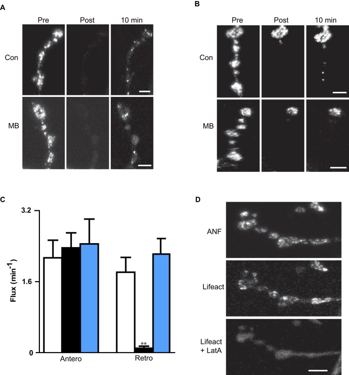
FIGURE 4:
MB inhibits retrograde but not anterograde DCV transport in the nerve terminal. (A) FRAP shows that nerve terminal anterograde DCV transport is maintained in MB. ANF-GFP images from the intact neuromuscular junction are oriented with the most distal bouton at the top. Scale bar, 3 μm. (B) SPAIM shows that retrograde transport out of the most distal bouton (at top of images) is blocked by MB. Scale bar, 3 μm. (C) Quantification from time-lapse experiments of anterograde DCV flux in the terminal and retrograde DCV flux out of the most distal bouton in controls (Con, open bars, n = 8), MB (black bars, n = 5), and LatA (blue bars, n = 4). **p < 0.01. (D) Lifeact-Ruby in terminal before and after treatment with LatA. Scale bar, 5 μm. ANF-GFP indicates the presence of DCVs.

