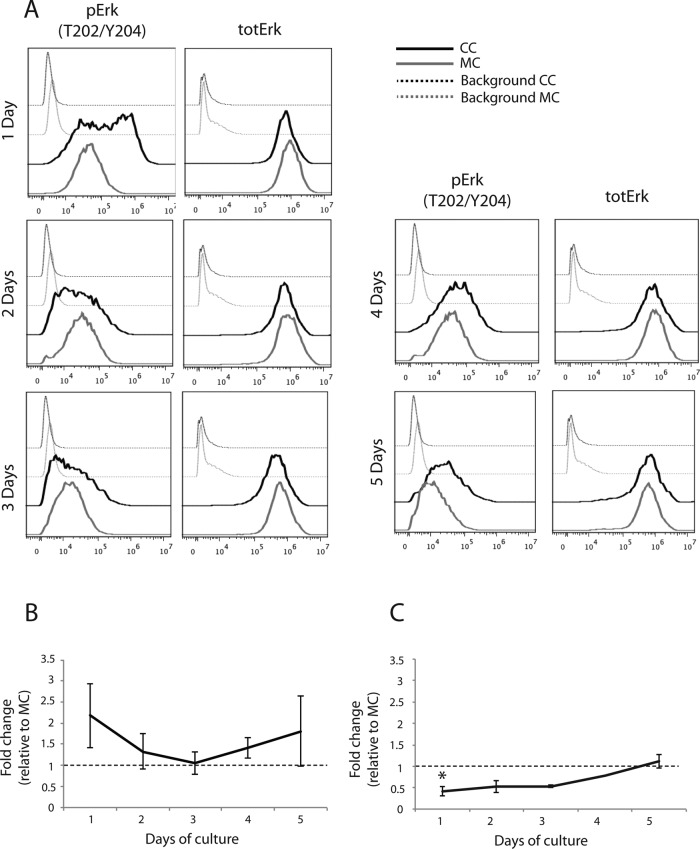
FIGURE 6:
Erk phosphorylation is up-regulated in ECs cocultured with vSMCs. We used three-color flow cytometry to investigate phosphorylation of Erk and total Erk expression in cocultured HUVECs. Erk phosphorylation was strongly up-regulated in the initial 2 d of coculture compared with monocultured HUVECs (A), as confirmed by quantification of relative geometric mean of fluorescence from two to four individual experiments (B). Total Erk was slightly reduced in cocultured HUVECs during the initial 3 d of coculture compared with monocultured HUVECs, although this reduction was only significant at 24 h (A, C). Asterisk indicates statistically significant differences from control (set to 1). *p < 0.05, min n = 2–4, except for C, 4 d, for which n = 1.

