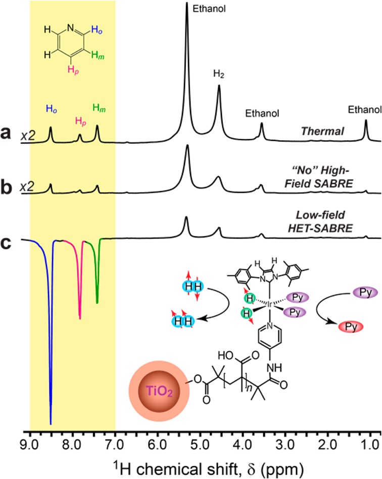
Figure 5.

(a) 1H NMR spectrum from a mixture containing d6-ethanol solvent, the nanoSABRE catalyst particles (NPCs), and the (fully protonated) pyridine substrate thermally polarized at 9.4 T following activation with pH2 bubbling. (b) 1H NMR spectrum of the sample in (a) obtained after nanoSABRE catalyst activation but with pH2 bubbling occurring at high field, acquired immediately after cessation of pH2 gas bubbling; no high-field (in situ) SABRE effect was observed. (c) 1H (ex situ) HET-SABRE NMR spectrum obtained from the same sample, acquired immediately after 30 s of pH2 bubbling at low field (∼100 G) and rapid transfer of the sample into the NMR magnet. All spectra shown were acquired with a single scan (90° pulse). Peaks at about δ ≈1.1, ≈3.6, and ≈5.2 ppm are from residual protons from the deuterated ethanol solvent. The peak at ≈4.5 ppm is from oH2. The inset shows the expected hexacoordinate structure of the activated catalytic moiety, exchanging with pH2 and the substrate (Py).
