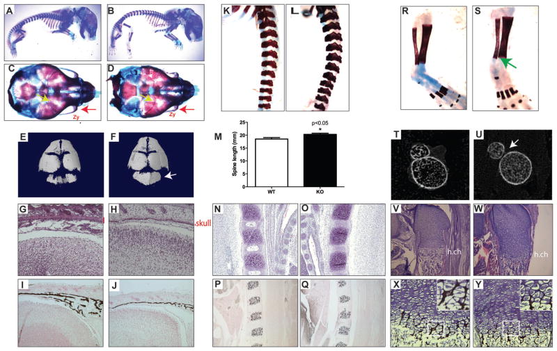
Fig. 6.
GATA4 cKO mice have skeletal defects. (A–D) Skeletal preparations of E18.5 WT (A, C) and cKO (B, D) mice. (A, B) Whole body. (C, D) Superior view of skull. Red arrow indicates decreased zygomatic bone size in cKO mice. Yellow arrows highlight a suture defect in the cKO skull. (E, F) μCT images of WT (E) and cKO (F) skulls. Arrow points to occipital bone. (G, H) H&E staining of sagittal sections of WT (G) and cKO (H) heads. Red line indicates thickness of skull bone. (I, J) Von Kossa staining of sagittal sections of WT (I) and cKO (J) heads. (K, L) Skeletal preparations of spine from WT (K) and cKO (L). (M) Total spine length of n = 7 mice. *p < 0.05. (N, O) H&E staining of sagittal sections of WT (N) and cKO (O) vertebrae. (P, O) Von Kossa staining of sagittal sections of WT (P) and cKO (Q) vertebrae. (R, S) Skeletal preparations of tibia and fibula from WT (R) and cKO (S). Green arrow indicates lack of fusion of tibia and fibula. (T, U) μCT images of WT (T) and ckO (U) tibia and fibula at their closest distance. (V, W) H&E staining of sagittal sections of WT (V) and cKO (W) femurs. (X, Y) Von Kossa staining of sagittal sections of WT (X) and cKO (Y) femurs. Zy = zygomatic bone; h.ch = hypertrophic chondrocytes. KO = knockout; cKO = conditional knockout.

