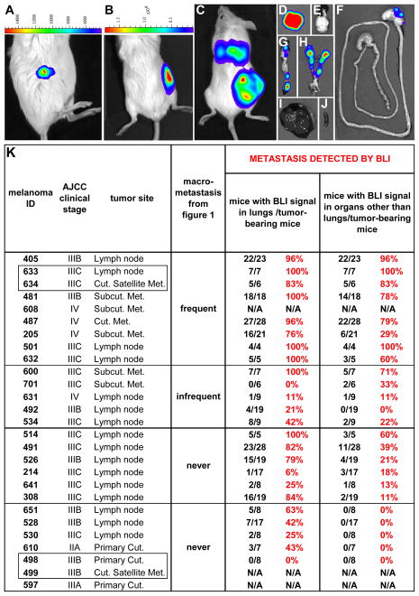
Figure 2. Bioluminescence imaging (BLI) confirms intrinsic differences in metastatic efficiency among human melanomas in NSG mice.
Luciferase-GFP+ melanoma cells from different patients exhibited intrinsic differences in metastatic efficiency revealed by BLI in NSG mice. BLI of an NSG mouse 2 days (A) and 45 days (B) after subcutaneous transplantation of 100 luciferase-GFP+ cells from melanoma 205, a heavily pigmented melanoma (see Fig. 1B). (C) BLI of an NSG mouse 13 weeks after subcutaneous transplantation of 100 luciferase-GFP+ cells from melanoma 487. BLI of individual organs dissected from the mouse in (C) revealed metastases in the lungs (D), stomach (F), pancreas (G), kidneys, adrenal glands and ovaries (H), but not in the brain (E), liver (I) or spleen (J). The maximum luminescence shown in red in panels C and D is 31x106 photons/second/cm2/steradian and in panels E–J is 6.2x10⁶ photons/second/cm2/steradian. (K) A summary for each melanoma of the percentage of mice with subcutaneous tumors that developed metastases detected by BLI. Supplementary Table 4 summarizes the locations in which metastases were detected by BLI. Melanomas 608, 499, and 597 did not undergo BLI.

