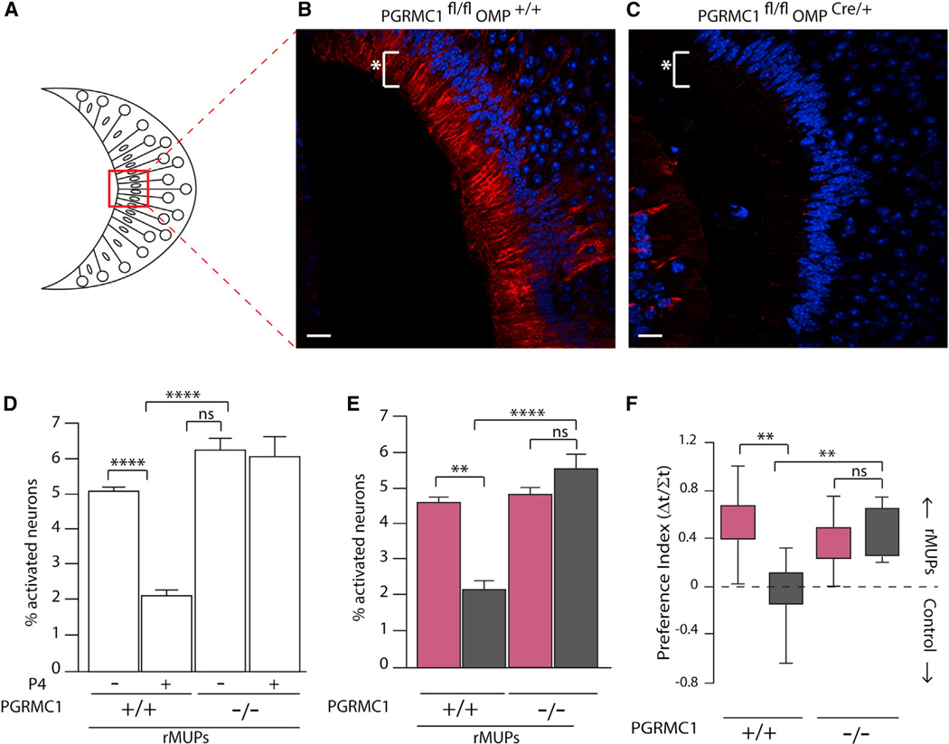
Figure 3. Silencing of VSN Activity by Progesterone Requires PGRMC1.
(A–C) Schematic of coronal VNO epithelium to orient (B) and (C). Immunohistochemical staining for PGRMC1 in (B) Pgrmc1fl/flOmp+/+ and (C) Pgrmc1fl/flOmpCre/+ mice. Scale bar, 20 µm, white asterisk indicates VSN dendrites.
(D)Percentage of VSNs showing calcium influx to rMUPs from ovx Pgrmc1fl/flOmp+/+ and Pgrmc1fl/flOmpCre/+ females treated with or without 40 nM P4 (2,112; 2,041; 2,077; and 2,085 cells imaged, respectively).
(E) Percentage of VSNs showing calcium influx to rMUPs from estrous and diestrous Pgrmc1fl/flOmp+/+ and Pgrmc1fl/fl OmpCre/+ females (2,543; 2,039; 2,549; and 2,223 cells imaged, respectively).
(F) Preference index from two choice behavior assay conducted on estrous and diestrous Pgrmc1fl/flOmp+/+ and Pgrmc1mlOmpCre/+ females (n = 8, 9, 8, and 8, respectively).
(D–F) One-way ANOVA followed by Bonferroni correction. All values in mean ±SEM. *p <0.05, **p< 0.01, ***p< 0.001, ****p< 0.0001; ns, not significant. White bars, ovx; pink bars, estrous; dark gray bars, diestrus.
See also Figure S3.

