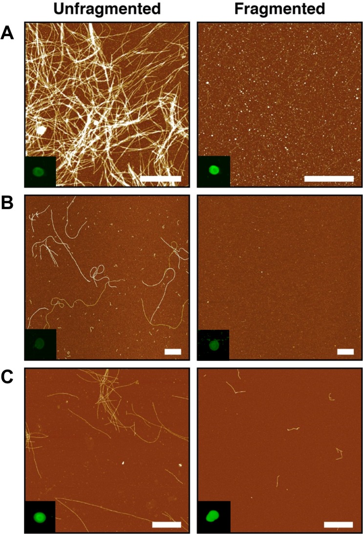
Fig 1. Amyloid fibril characterization.

AFM height images of fibril samples adhered to mica surface (scale bar equals 1 μm) (A) α-syn, (B) Lysozyme and (C) Aβ40 with dot blot analysis of fibril samples (inset) by anti-fibril LOC antibody specific to generic epitopes common in amyloid fibrils.
