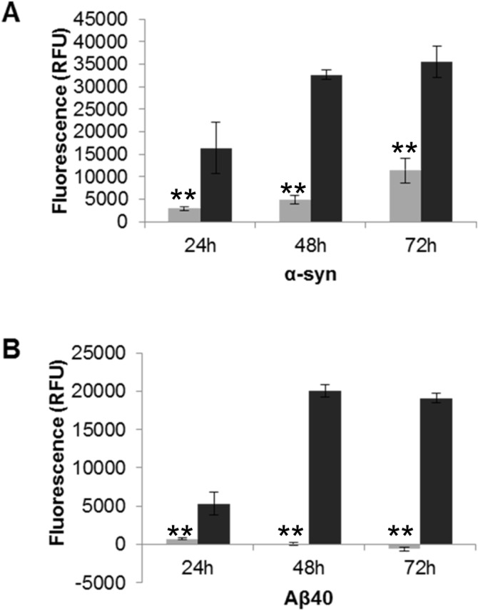
Fig 3. The effect of fragmented fibril on cell viability by cell membrane integrity assay.

SH-SY5Y cells (2x10⁴/well) were plated with the addition of the CellTox dye and allowed to adhere. The unfragmented and fragmented fibril samples were then added (A) α-syn 7 μM and (B) Aβ40 10 μM. Fluorescence was recorded over a period of 72 hours (520Em/ 485Ex). Light grey bars denote unfragmented fibril samples and dark grey bars denote fragmented fibril samples (n = 3, error bars show SE). Unfragmented (light grey) and fragmented fibril samples (dark grey) (n = 6, SE). Statistical significance between toxicity of unfragmented and fragmented fibrils at each time point was determined by a Mann Whitney U test P ≤ 0.05 (*) or P ≤ 0.005 (**).
