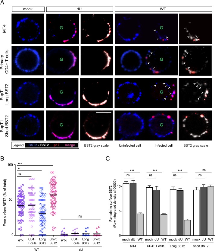
Fig 6. Residual BST2 clusters are detected outside virus assembly sites in the presence of Vpu.
MT4, primary CD4+ T cells, SupT1-shortBST2 and SupT1-longBST2 cells were mock-infected (mock) or infected with VSV-G-pseudotyped NL4.3-Ada-GFP WT or dU viruses. (A) Cells were stained with anti-BST2 Abs (blue), fixed, permeabilized and then sequentially stained with anti-p17 Abs (red). Infected cells (GFP+) are marked with a green letter G. An uninfected cell is shown next to WT-infected cells as indicated. Clusters of free BST2 are marked with white open arrows. White bar = 10 μm. (B) The number of residual BST2 clusters not co-localizing with p17 (designated as free BST2) per cell was calculated and expressed as the percentage of the total number of surface BST2 clusters. (C) Quantitative analysis of surface BST2 was determined as described in Materials and Methods. One way ANOVA with Bonferroni’s multiple comparison test was used (*** p<0.001, ** p<0.01, ns not significant (p>0.05)). Error bars indicate the standard error of the mean after analysis of at least 50 distinct cells.

