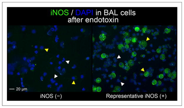
FIGURE 2.

Immunohistochemical staining for iNOS (green) in cells obtained by BAL in endotoxin-challenged airway. Only 1 individual had negative iNOS staining (iNOS (−)). iNOS (+) image is representative of positive staining results obtained on BAL cells from 6 volunteers. Neutrophils (white arrowheads) and macrophages (yellow arrowheads) were identified by nuclear morphology from 4′,6-diamidino-2-phenylindole staining (blue). Images taken at ×20 magnification.
