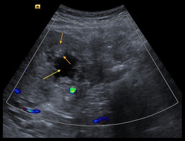
Figure 1.

Trans-abdominal ultrasound image with color flow demonstrates an irregular, thick-walled (between orange arrows) mass in the region of the gallbladder fossa with cystic/necrotic center (yellow arrow). The wall of the mass lacks increased vascularity.
