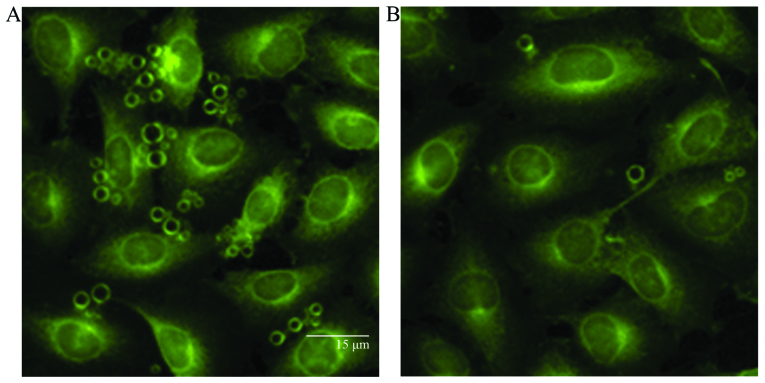
Figure 2.
Combination of targeted microbubbles with ECV304 cells stimulated or not with interleukin-1β (IL-1β) as observed under a fluorescence microscope (x200 magnification). (A) The fringe of the IL-1β stimulated ECV304 cells showed bright green fluorescence. (B) Slight green fluorescence was observed at the fringe of the normal ECV304 cells not stimulated with IL-1β.

